Micro Topography Patterning of General Elastomer Surfaces for the Endothelialization of Ventricular Assist Devices
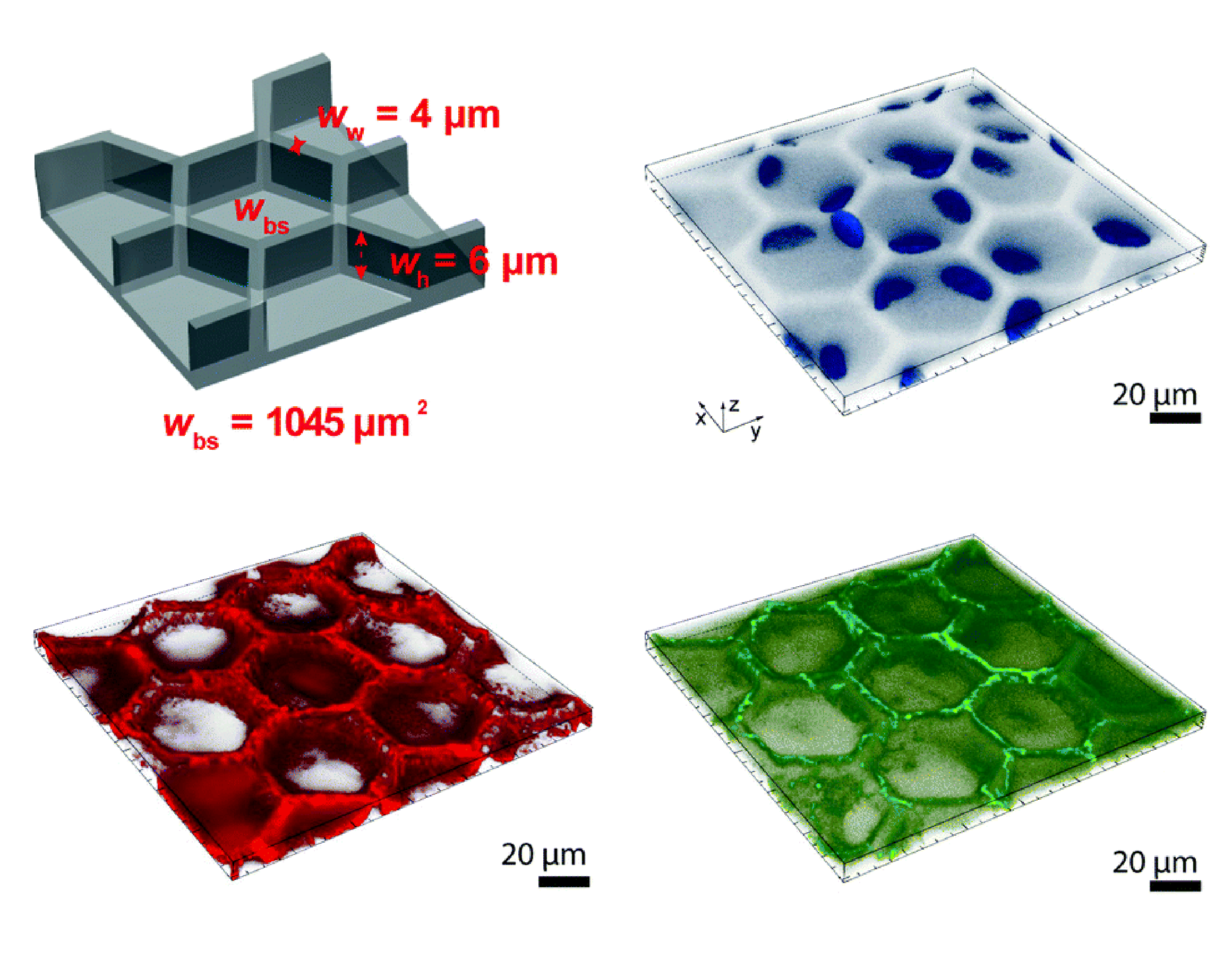
Synthetic substrate with a monolayer of endothelial cells (EC) could form the basis of a 100% haemocompatible blood pump. However, cells could experience supra-physiological wall shear stress (WSS) in artificial blood pumps such as Ventricular assist devices (VADs). This supra-physiological mechanical signal that cells sense would discourage their stay on the substrate and cause monolayer disassembly. In order to maintain an EC monolayer under supra-physiological WSS, an array of Hexagonal wells [1], whose repeating unit showed in Figure 1 (left), was developed to protect cells under such conditions. Human umbilical vein EC (HUVEC) were able to hide under the well when exposed to supra-physiological WSS, as shown in Figure 1 (right). However, its fabrication process with soft lithography makes it difficult to apply such technology on complex geometries, such as curved surfaces.
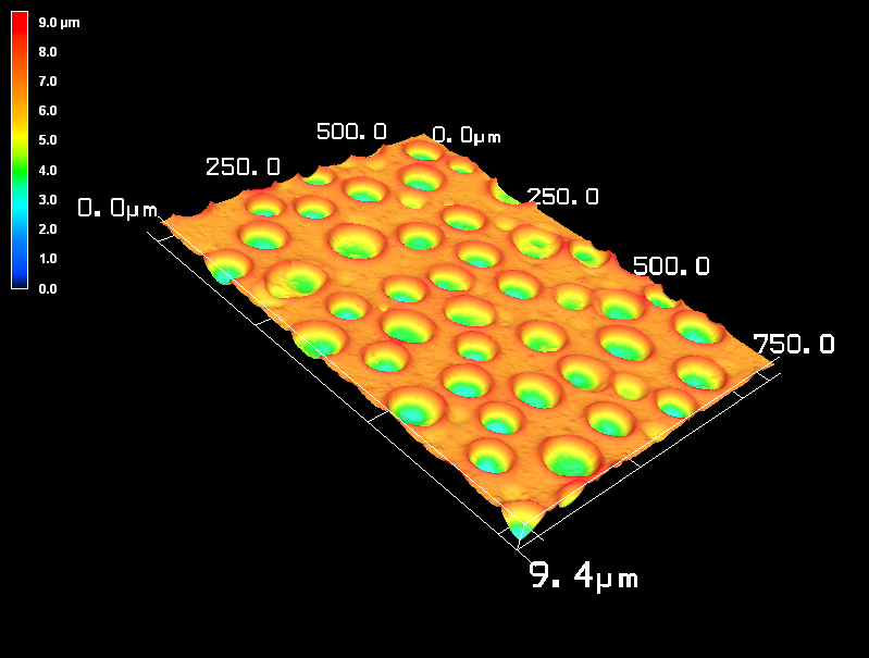
In this project, a novel method called ‘Breath Figures’ (BF) is developed to create similar topography. This method utilizes the nucleation of microscale water droplets on a deformable substrate to form microscale dimples. This self-assembly method is tunable and applicable on different surfaces. A 3D characterization of such surface with the BF topography is shown on Figure 2.
Static and dynamic cell cultures are then performed on the substrate with the BF topography. EC formed a connected monolayer successfully under static environment on the topography with a depth up to 10 microns. A custom-built laminar flow chamber [2] were used to verify monolayer integrity under differently conditions of WSS. Under changing flow direction and changing WSS magnitude, better overall monolayer integrity was observed on the BF patterned surface. The better monolayer integrity was associated with a less adaptation to specific flow conditions. Furthermore, this difference in monolayer response was attribute primarily to the differenced in VE-Cadherin phosphorylation on Tyrosine 658.
Details of the work has been pubslished on Biomaterials [3] in 2021. The BF technology was also filed under European Patent No. EP20208958.7 and International Patent PCT/EP2021/081325.
This work is part of the Zurich Heart project under the umbrella of University Medicine Zurich/Hochschulmedizin Zürich.
Project Lead

Xi Wu
[1] B.J. Bachmann, C. Giampietro, A. Bayram, G. Stefopoulos, C. Michos, G. Graeber, M.V. Falk, D. Poulikakos, A. Ferrari, Honeycomb-structured metasurfaces for the adaptive nesting of endothelial cells under hemodynamic loads, Biomater. Sci. 6 (2018) 2726–2737.
[2] D. Franco, F. Milde, M. Klingauf, F. Orsenigo, E. Dejana, D. Poulikakos, M. Cecchini, P. Koumoutsakos, A. Ferrari, V. Kurtcuoglu, Accelerated endothelial wound healing on microstructured substrates under flow, Biomaterials. 34 (2013) 1488–1497. https://doi.org/10.1016/J.BIOMATERIALS.2012.10.007.
[3] X. Wu, S. Moimas, R. Hopf, C. Giampietro, A. Kourouklis, V. Falk, E. Mazza, A. Ferrari, A free-form patterning method enabling endothelialization under dynamic flow, Biomaterials. 273 (2021) 120816. https://doi.org/10.1016/J.BIOMATERIALS.2021.120816.