Skin Multilayer Mechanobiology
A layered mechanical model of the skin
Skin is a complex structure composed of several distinct layers. In general, as depicted in figure 1, the three main layers accounted for are epidermis, dermis and hypodermis. Each of them consists of different types of cells and fulfills different functions while interacting with our environment and protecting us against external threats. The epidermis, the outermost layer can further be subdivided into several strata starting from the stratum corneum, the final layer of the skin, and ending in the basement membrane. The basement membrane connect epidermis and dermis. Due to its relative size and its high collagen content the dermis is generally considered as the main load bearing layer of the skin in extension. While its top part, the papillary dermis, consist of fine, densely packed collagen fibers, the lower part, the reticular dermis, is primarily build of thicker collagen bundles in a wavy arrangement. Below the dermis, there is the hypodermis, a space for energy storage and a protection against cold temperatures and external pressure to our body. This layer mainly consists of adipocytes hold together by the interlobular septa, once again a collagenous structure.
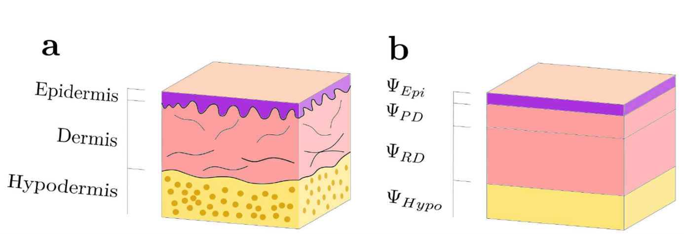
Traditionally, empirical skin modeling approaches assume an incompressible single-phase material. Moreover, they often do not differentiate between the aforementioned different layers. More recently however, experimental results suggest to model skin as well as other soft biological tissues as multiphasic materials formed of a matrix material, a fluid and several ions.
Based on these new findings we will propose a multiphasic model of skin. This model considers its different phases and accounts for its layered structure. Special consideration is taken to utilize this model to reliably predict stresses and strains within the different layers as well as characterizing the osmotic pressure and the resulting interstitial flow.
Mechanosensitive Processes in Skin
Mechanical forces can alter biological processes and especially growth and wound healing. A practical example is the direction of cuts made by surgeons. In order to reduce tension on wounds, skin is cut parallel to Langer lines and not perpendicular. Looking at smaller length scales, skin cells possess mechanosensitive elements, which trigger processes, such as growth. Especially osmotic pressure, hydrostatic pressure, the tissue stiffness and the fluid shear stress is “felt” by cells and resulting in the cell adapting to the new “mechanical homeostasis”.
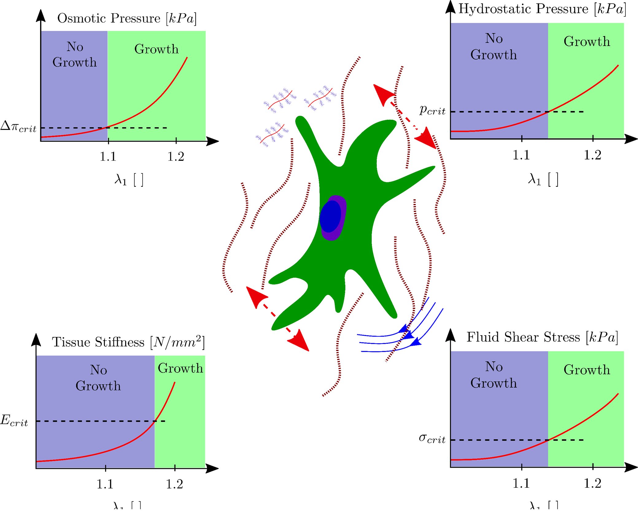
Modeling and predicting mechanobiological processes requires simulations taking into account evolution equations for the mechanical forces, the biological processes as well as their interactions. Recently so called "system based approaches", combining all those evolution equations, have emerged. This project will develop suitable computational tools to study the mechanobiology of skin.
This work is conducted as part of external page the SKINTEGRITY flagship project of University Medicine Zurich and financially supported by the Swiss National Science Foundation (Grant No. 179012: “Skin biomechanics and mechanobiology for wound healing and tissue engineering”).
Project Lead

David Sachs