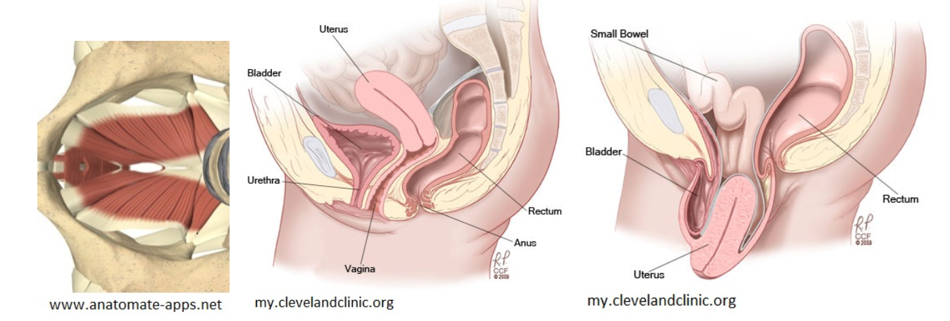
3D Finite Element Pelvic Floor Modeling
Pelvic floor dysfunctions affect a wide range of women of all age groups, more than 10% of which require surgery. However the mechanisms resulting in such pathologies are still poorly understood.


In order to get a better understanding of the patient specific factors leading to pelvic floor weakness, we propose a new diagnostic tool conceptualized in a virtual 3D finite element model of the pelvic region.
This was built based on a finite element mesh of a segmented geometry from MRI data of a pregnant healthy woman, provided by the group of Prof. Brieu in Lille, France. It consists of the pelvic floor muscles, bladder, vaginal canal/uterus and several important ligaments. The applied boundary conditions are representative of physiological conditions.

Current methods to evaluate pelvic region kinematics in vivo (under MRI or ultra sound) consist mostly of applying a downwards pressure by either the patient itself (intra-abdominal pressure) or by pushing externally on the belly region. This is rather inconsistent and cannot be precisely mechanically controlled. We develop a new procedure with which intra vaginal boundary conditions are applied and controlled precisely. In a preliminary study the influence of the interface between the bladder and vaginal canal on the kinematics of the pelvic system has been investigated. This interface (fascias and connective tissue) was included as an individual model part with adjustable mechanical characteristics (see images, soft interface on the left, hard interface on the right).
Project Lead
Manfred Maurer
Partners
Prof. Brieu, Laboratoire de Mécanique de Lille, France
Prof. Dr. Jan Deprest, Katholieke Universiteit Leuven, Belgium
Dr. David Scheiner, Department of Gynecology, University Hospital Zurich
Prof. Dr. Caroline Maake, Institute of Anatomy, University of Zurich