Microstructural Model of Fetal Membranes

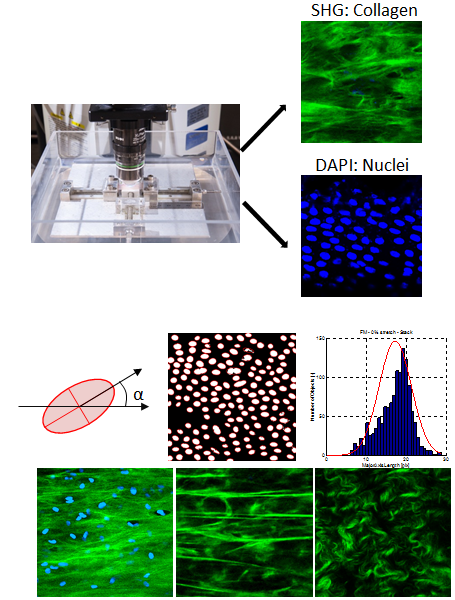
The mechanical response of fetal membranes shows particular features related to their function: very compliant response in uniaxial stress state, large biaxial stiffness, large transversal contraction and multilayer character. In order to better understand the mechanisms of deformation underlying the tissue microstructure, a new stretching device is used for in situ measurements in a multiphoton microscope. This set up allows to visualize the whole thickness of the membranes and quantitatively characterize modifications of the microstructure (fiber orientation) under different loading conditions. The second harmonic generation of the collagen fiber network and the fluorescence of the nuclei (DAPI staining) are acquired simultaneously in two different channels.

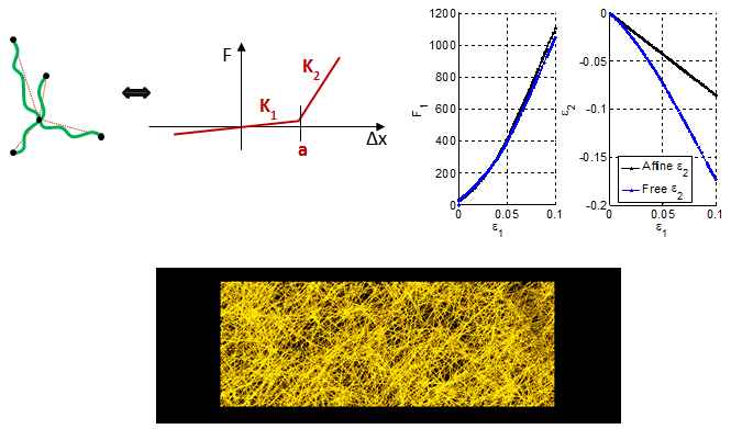
Tissue microstructure is considered in the mechanical model, following two approaches: (i) In a random fibers network model, based on nonlinear connectors. Parameters such as fibers density, interconnectivity, non-linear force law are evaluated with respect to the response of a RVE subjected to the loading conditions applied in the experiments. (ii) A continuum model (akin to the formulation of Gasser et al. 2006) including a fiber family, which is described by a mean direction and a fiber dispersion. These two parameters and their evolution can be linked to the observed fiber orientation.
Project Lead
Arabella Mauri
Partners
Prof. Zimmermann, USZ, Obstetrics and Gynecology
Prof. Maake, Institute of Anatomy, University of Zurich
Dr. José Maria Mateos, Center for Microscopy and Image Analysis, University of Zurich
Funding
Swiss National Science Foundation (SNSF)